Weak bone density in the spine may indicate a decline in cognitive abilities
According to the latest data obtained, low bone mineral density in the thoracic spine is associated with a faster decline in cognitive functions and accelerated damage to the brain’s white matter, which is characteristic of aging. The study is based on a secondary analysis of data from the large‑scale Multi‑Ethnic Study of Atherosclerosis (MESA). The final sample included 715 participants for whom volumetric bone mineral density (vBMD) measurements in the thoracic vertebrae, brain MRI results, and cognitive test data were available.
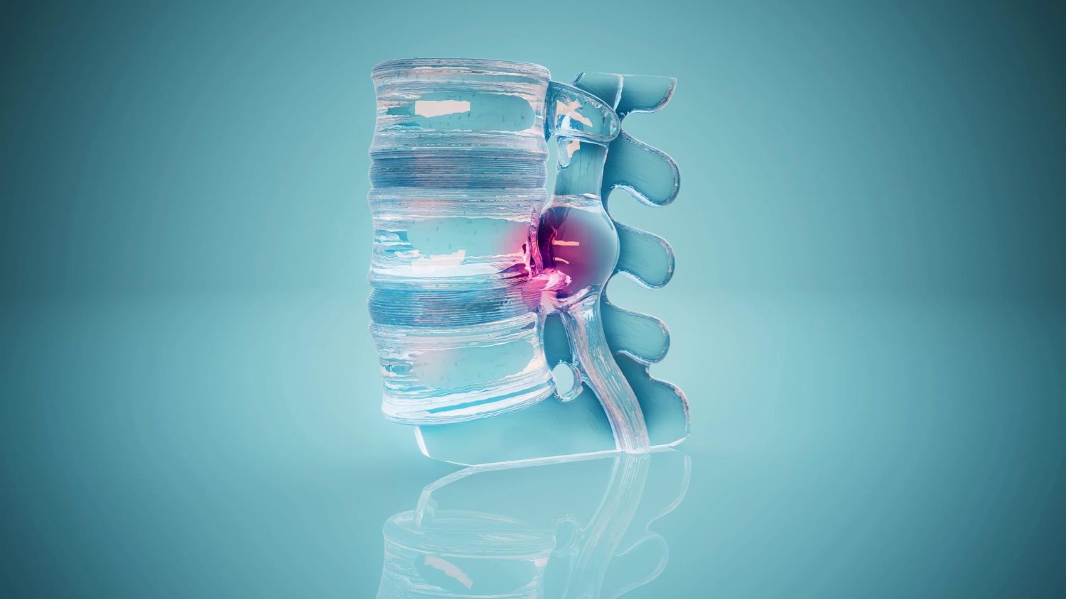
Bone density was calculated using an algorithm based on artificial intelligence — it processed conventional non‑contrast CT scans of the chest. This approach made it possible to extract additional information from existing studies that are usually performed for other indications, for example, for lung cancer screening or monitoring nodules.
The key finding was that low initial bone density correlated with an accelerated decline in overall cognitive performance. Moreover, patterns were observed in specific areas of the brain. Thus, lower bone density was associated with the accumulation of white matter hyperintensities in the corpus callosum — a structure responsible for executive functions, working memory, and attention. A more pronounced decrease in fractional anisotropy was also identified in the anterior limb of the internal capsule, another area important for planning and controlling actions. These MRI changes are considered markers of age‑related brain damage and are associated with an increased risk of dementia.
Low bone density itself does not cause cognitive decline. Rather, this is a reflection of the general mechanisms of aging that simultaneously affect both the skeleton and the brain. Such common factors may include, for example, insulin resistance, lipid metabolism disorders, or hormonal changes during menopause.
The practical value of this data lies in the fact that it provides a simple way to identify people at risk in advance. Since chest CT scans are performed quite frequently, doctors have the opportunity to simultaneously assess bone density in the vertebrae and, based on this, suggest earlier monitoring of cognitive health and correction of common risk factors.
According to Dr. Shadpour Demeri, modern technologies make it possible to extract much more data from diagnostic images than before. Artificial intelligence is capable of analyzing images across several organ systems simultaneously and identifying relationships between age‑related pathologies that were previously studied in isolation.
Published
September, 2026
Updated
Category
Medicine
Duration of reading
2-3 min
Share


Source
Scientific journal Radiology. Article: Deep Learning-derived Bone Mineral Density and Longitudinal White Matter Microstructure and Cognitive Decline: Multi-Ethnic Study of Atherosclerosis
Global health science — in clear language
We select and translate the best international publications. Read 3 key materials about longevity every week!