Scientists have created a living intervertebral disc to combat back pain
The UK has a new tool that could revolutionize the approach to one of the world’s most common problems – chronic back pain. A team of experts from the University of Manchester has developed a full-fledged model of a human intervertebral disc using bioprinting technology.
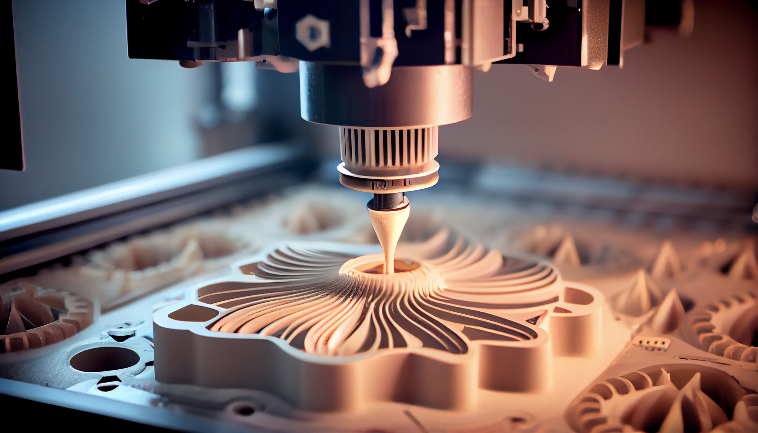
The technology is based on 3D printing, but instead of plastic, it uses living cells and gel-like materials such as collagen and alginate, a substance derived from seaweed. This makes it possible to recreate not just the shape of the organ, but also its internal environment, chemical and mechanical properties.
According to Dr. Matthew Kibble, the author of the project, the team was able to print a disc as close to the real thing as possible – with the same structure, cell types and level of tissue density. The model allows them to observe exactly how back pain occurs at the cellular level and how the disc tissue deteriorates over time.
Two key factors were emphasized – tissue stiffness and oxygen levels. These parameters directly affect the production of important components such as collagen and hyaluronic acid. Both substances play a crucial role in maintaining spinal health, and disruption of their synthesis can lead to pain, inflammation and disc degeneration.
The work utilized advanced equipment capable of layering different cell types and materials. The resulting biomodels were placed in specially controlled conditions so that they “matured” and began to function like real tissue.
Dr. Stephen Richardson, co-author of the project, noted that this discovery is a step toward creating realistic models of organs and a deeper understanding of the causes of disc destruction. This approach offers hope for more accurate and effective ways of tissue repair, including the use of stem cells and gene therapy.
Bioprinting technology has already been used to create models of skin, brain, heart and other organs. For now, however, such designs serve more for studying processes in the laboratory and partially replace experiments on animals.
According to the team, the next step will be to integrate cells from healthy young discs and stem cells to create even more complex and viable models. This will not only allow them to understand how healthy tissue forms, but also possibly create complete replacements for destroyed discs in the future.
Millions of people around the world suffer from low back pain. A new approach that allows functional models of discs to be printed could be the game changer that leads to breakthrough treatments.
Published
May, 2025
Duration of reading
2-3 minutes
Category
New technologies
Share


Don’t miss the most important science and health updates!
Subscribe to our newsletter and get the most important news straight to your inbox