Structure and function of joints
A joint is a movable joint of two or more bones of the skeleton.
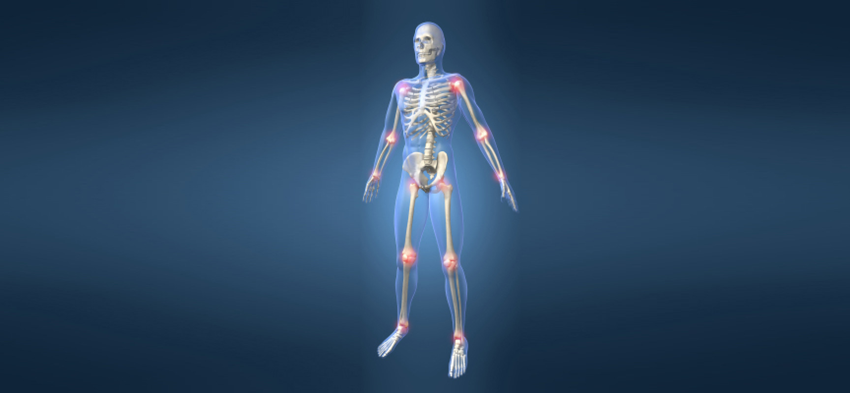
Joints unite the bones of the skeleton into a single whole. More than 180 different joints help a person move. Together with bones and ligaments, they are classified as the passive part of the musculoskeletal system. Joints can be compared to hinges, whose task is to ensure smooth sliding of bones relative to each other. In their absence, the bones will simply rub against each other, gradually collapsing, which is a very painful and dangerous process. In the human body, joints play a threefold role: they help maintain body position, are involved in moving body parts relative to each other, and are organs of locomotion (movement) of the body in space.
The main elements that are present in all so-called true joints are:
- articular surfaces (ends) of connecting bones;
- joint capsule;
- joint cavity.
The joint cavity is filled with synovial fluid, which is a kind of lubricant and promotes the free movement of the articular ends.
According to the number of articular surfaces, there are:
- a simple joint with only 2 articular surfaces, such as the interphalangeal joints;
- a complex joint with more than two articulating surfaces, such as an elbow joint. A
- complex joint consists of several simple joints in which movements can be performed separately;
- a complex joint containing intra-articular cartilage that divides the joint into 2 chambers (a two-chamber joint).
The classification of joints is carried out according to the following principles:
- by the number of articular surfaces;
- by the shape of the articular surfaces;
- by function.
The articular surface of the bone is formed by hyaline (less often fibrous) articular cartilage. Articular cartilage is a tissue filled with fluid. The cartilage surface is smooth, strong and elastic, capable of absorbing and releasing fluid well. The thickness of articular cartilage averages 0.2-0.5 millimeters.
The joint capsule is formed by connective tissue. It surrounds the articulating ends of the bones and passes into the periosteum on the articular surfaces. The capsule has a thick outer fibrous fibrinous membrane and an inner thin synovial membrane that secretes synovial fluid into the joint cavity. The ligaments and tendons of the muscles strengthen the capsule and promote joint movement in certain directions.
The auxiliary formations of the joint include intraarticular cartilage, discs, menisci, lips and intracapsular ligaments. The blood supply to the joint is carried out from a widely anastomosing (branched) articular arterial network formed by 3-8 arteries. The innervation (supply of nerves) of the joint is carried out by a nervous network formed by sympathetic and spinal nerves. All articular elements, except hyaline cartilage, have innervation. They contain significant amounts of nerve endings that carry out pain perception, as a result of which they can become a source of pain.
Joints are usually divided into 3 groups:
- synarthrosis — fixed (fixed);
- amphiarthrosis (semi—articular) – partially mobile;
- diarthrosis (true joints) are movable. Most joints are movable joints.
According to the World Health Organization, every 7th inhabitant of the planet suffers from joint pain. Between the ages of 40 and 70, joint diseases are observed in 50% of people and in 90% of people over 70 years of age.
A synovial joint is a joint where the ends of the bones meet in a joint pouch. These include most human joints, including the supporting knee and hip joints.
Joints are divided into simple and complex. The formation of simple bones involves 2 bones, complex bones — more than 2 bones. If several independent joints are involved in the movement, as in the lower jaw when chewing, such joints are called combined. A combined joint is a combination of several isolated joints located separately but functioning together. These are, for example, both temporomandibular joints, the proximal and distal radiocarpal joints, and others.
In shape, the articular surfaces resemble segments of the surfaces of geometric bodies: a cylinder, an ellipse, a ball. Depending on this, cylindrical, ellipsoid and spherical joints are distinguished.
The shape of the articular surfaces determines the volume and direction of movements around the 3 axes: sagittal (runs from front to back), frontal (runs parallel to the plane of the support) and vertical (perpendicular to the plane of the support).
Circular motion is a sequential motion around all axes. In this case, one end of the bone describes a circle, and the whole bone forms a cone shape. Sliding movements of the articular surfaces are also possible, as well as their removal from each other, as is observed, for example, when stretching the fingers. The function of a joint is determined by the number of axes around which movements are performed.
There are the following main types of joint movements:
- movement around the frontal axis — flexion and extension;
- movements around the sagittal axis are the reduction and withdrawal of movement around the vertical axis, that is, rotation: inward (pronation) and outward (supination).
The human hand contains: 27 bones, 29 joints, 123 ligaments, 48 nerves and 30 named arteries. Throughout our lives, we make finger movements millions of times. The movement of the hand and fingers is provided by 34 muscles, only 9 different muscles are involved in the movement of the thumb.
Don’t miss the most important science and health updates!
Subscribe to our newsletter and get the most important news straight to your inbox
Shoulder joint
It is the most mobile in humans and is formed by the head of the humerus and the articular cavity of the scapula.
The articular surface of the scapula is surrounded by a ring of fibrous cartilage — the so-called articular lip. The tendon of the long head of the biceps brachii passes through the joint cavity. The shoulder joint is strengthened by the powerful coracoid—shoulder ligament and surrounding muscles – deltoid, scapular, supraspinatus and subclavian, large and small round. The large pectoral and latissimus dorsi muscles also participate in shoulder movements.
The synovial membrane of the thin articular capsule forms 2 extra—articular turns – the tendons of the biceps brachii and the scapular muscles. The anterior and posterior arteries encircling the humerus and the thoracoacromial artery participate in the blood supply to this joint, venous outflow is carried out into the axillary vein. The outflow of lymph occurs in the lymph nodes of the axillary region. The shoulder joint is innervated by branches of the axillary nerve.
Movements around 3 axes are possible in the shoulder joint. Flexion is limited by the acromial and coracoid processes of the scapula, as well as the coracoid-humeral ligament, extension by the acromion, coracoid-humeral ligament and joint capsule. Abduction in the joint is possible up to 90 °, and with the participation of the upper limb girdle (with the inclusion of the sternoclavicular joint) — up to 180 °. The abduction stops at the moment when the large humeral humerus stops in the coracoid-acromial ligament. The spherical shape of the articular surface allows a person to raise his arm, pull it back, rotate the shoulder along with the forearm, hand in and out. Such a variety of hand movements has become a crucial step in the process of human evolution. In most cases, the shoulder girdle and shoulder joint function as a single functional entity.
Hip joint
It is the most powerful and heavily loaded joint in the human body and is formed by the acetabulum of the pelvic bone and the head of the femur. The hip joint is reinforced by the intraarticular ligament of the femoral head, as well as the transverse ligament of the acetabulum, covering the neck of the femur. From the outside, the powerful ilio-femoral, pubic-femoral and sciatic-femoral ligaments are woven into the capsule.
blood supply to this joint is carried out through the arteries encircling the femur, by branches of the occlusive and (intermittently) branches of the superior perforating, gluteal and internal genital arteries. Blood outflow occurs through the veins surrounding the femur, into the femoral vein and through the occlusal veins into the iliac vein. Lymph outflow is carried out into the lymph nodes located around the external and internal iliac vessels. The hip joint is innervated by the femoral, occlusal, sciatic, superior and inferior gluteal and genital nerves.
The hip joint is a type of spherical joint. Movements around the frontal axis (flexion and extension), around the sagittal axis (abduction and adduction) and around the vertical axis (external and internal rotation) are possible in it.
This joint is under heavy strain, so it is not surprising that its lesions occupy the first place in the general pathology of the articular apparatus.
The knee joint
It is one of the largest and most complex joints in humans. It is formed by 3 bones: femoral, tibial and fibular. Intra- and extra-articular ligaments provide stability to the knee joint. The extraarticular ligaments of the joint are the fibular and tibial collateral ligaments, oblique and arched popliteal ligaments, patellar ligament, medial and lateral supporting ligaments of the patella. The intraarticular ligaments include the anterior and posterior cruciate ligaments.
The joint has many auxiliary elements, such as menisci, intra-articular ligaments, synovial folds, and synovial pouches. There are 2 menisci in each knee joint — an external and an internal one. The menisci have the appearance of half-moons and perform a cushioning role. The auxiliary elements of this joint include synovial folds, which are formed by the synovial membrane of the capsule. The knee joint also has several synovial pouches, some of which communicate with the joint cavity.
Everyone had to admire the performances of gymnasts and circus performers. People who can climb into small boxes and bend unnaturally are said to have gutta-percha joints. Of course, this is not the case. The authors of the Oxford Handbook of Body Organs assure readers that “such people’s joints are phenomenally flexible” — in medicine, this is called joint hypermobility syndrome.
The shape of the joint is a condyle joint. It allows movement around 2 axes: frontal and vertical (when bent at the joint). Flexion and extension occur around the frontal axis, and rotation occurs around the vertical axis.
The knee joint is very important for human movement. With each step, by bending, it allows the foot to step forward without hitting the ground. Otherwise, the leg would be brought forward by lifting the hip.
Source: Igor Platonov “Muscles and joints. Musculoskeletal system”
Published
July, 2024
Duration of reading
About 3-4 minutes
Category
Body
Share

